Flashes & Floaters

WHAT ARE FLOATERS?

Floaters are small particles that you see moving in your field of vision. They are in fact tiny areas of contracted vitreous; the clear jelly-like substance that fills the inside of the eye. We see them as spots, strands, webs or various other shapes because they cast shadows on the retina as light enters the eye. They are more noticeable when looking at a blank background such as a blue sky, a white wall or a computer screen.

WHAT CAUSES FLOATERS?

For most people, floaters occur naturally with age as the vitreous gel shrinks and changes consistency. As this occurs, it will eventually separate from the retina, causing a larger floater. Usually, this process occurs without any negative consequences, but in some cases, the gel pulls a piece of the retina with it, causing a hole or tear in the retina. If this goes untreated, fluid can collect under the retina causing it to detach. People who are highly near-sighted, diabetics and those who have experienced recent trauma to the eye are at greater risk of retinal detachment.

CAN FLOATERS BE TREATED?

If you suspect that you have floaters, be sure to schedule an appointment with one of our doctors to make sure that your condition isn’t serious. Floaters without any other symptoms such as crescent-like flashes of light are usually not problematic. If, however, you are also experiencing flashing lights in your peripheral vision or any sense of a curtain or shade encroaching from any direction, you could be having a more serious problem involving the retina that warrants immediate attention. Most floaters are harmless but annoying and they usually become less noticeable over time. Only in rare instances are floaters surgically removed.

Ocular Surface Disease

Ocular Surface Disease

Dry Eye Syndrome

Dry eye disease is a common condition that results from inadequate tear film composition. Your tear film is composed of 3 layers: aqueous fluid, oils from the meibomian glands and mucus. Some people don’t produce enough aqueous, some have increased evaporation from the surface and others produce a poor -quality tear film. This tear film instability leads to inflammation and damage to the surface of the eye which can significantly affect your quality of life.

What are the symptoms of dry eyes?

  • Burning, stinging, scratchy foreign body
  • Red eyes with stringy mucus
  • Light sensitivity
  • Excessive tearing: this may sound counterintuitive, but it is a reflexive response to dry eyes
  • Blurry vision that gets worse throughout the day
  • Difficulty wearing contact lenses

Who is at risk of developing Dry Eye Syndrome?

  • Women over 50, especially if experiencing hormonal changes due to pregnancy, menopause, or the use of birth control
  • People taking medication such as antihistamines, decongestants, anti-depressants, medications for high blood pressure, acne and chemotherapy
  • People with corneal nerve damage caused by contact lens use, previous laser eye surgery, infections and diabetes
  • People with eyelid disorders such as blepharitis or meibomian gland dysfunction, rosacea, entropion (eyelid turns in) and ectropion (eyelid turns out)

We offer comprehensive evaluation and management of all ocular surface disorders.

Systemic diseases

Systemic diseases

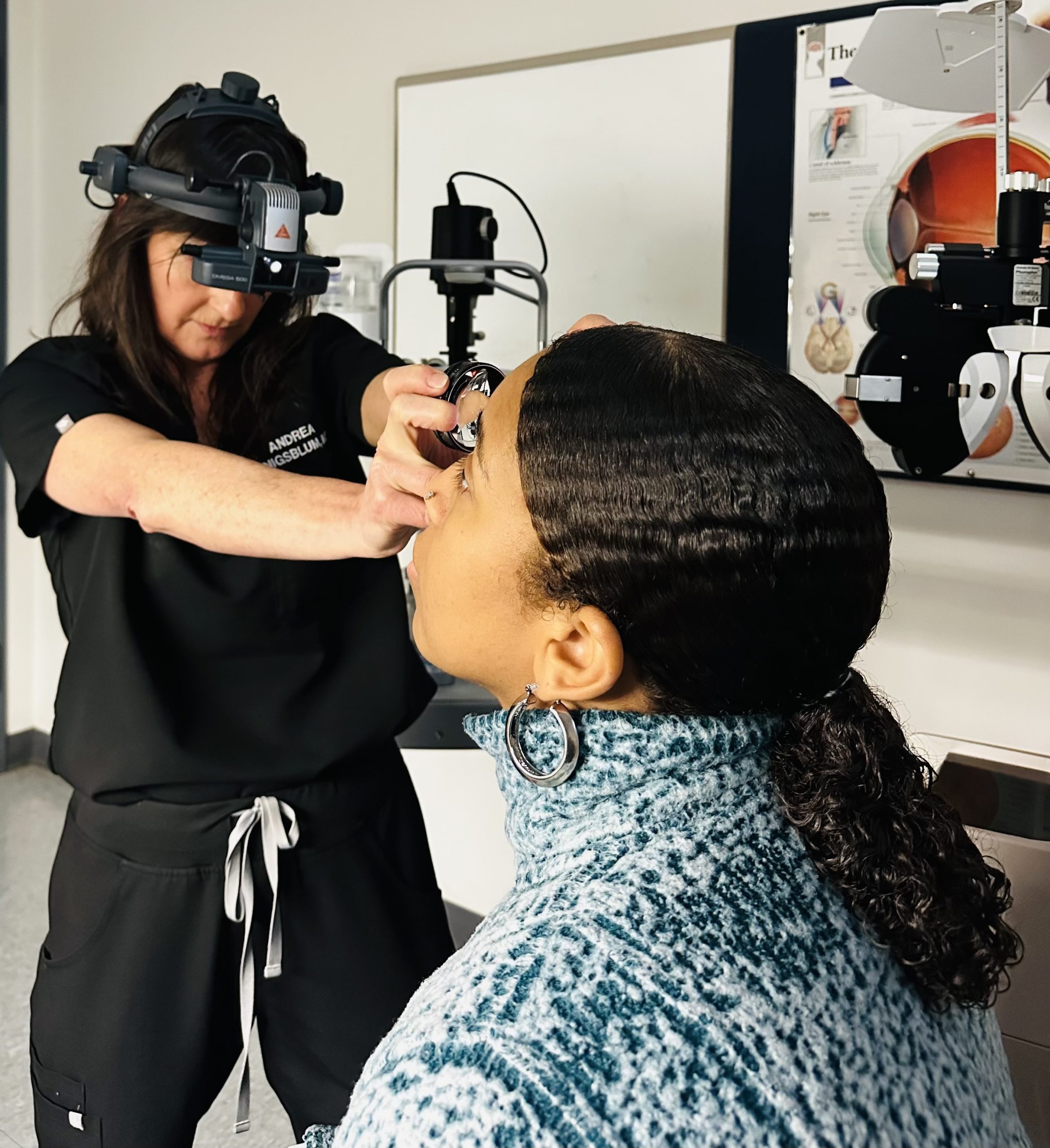
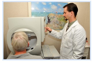
A comprehensive dilated eye exam by an ophthalmologist can detect a multitude of systemic diseases and is often the first indication of many disease processes. The retinal blood vessels are the only place where a doctor can directly view your circulation without the need for expensive invasive tests like angiograms, CT scans, or MRI. The eyes are truly the window through which we view the rest of the body.

What can my ophthalmologist diagnose from a dilated eye exam?

DIABETES

It is recommended that all diabetics have a dilated eye exam at least once a year. Pregnant women with gestational diabetes may require an exam every trimester.

Diabetes can cause transient blurry vision when first diagnosed due to swelling of your natural lens. This is often one of the first signs of elevated blood sugar. It can also cause a specific type of diabetic cataract causing significant glare and difficulty reading. Diabetes can even cause age related cataracts to progress at a faster pace. Diabetics are also at greater risk of developing glaucoma.

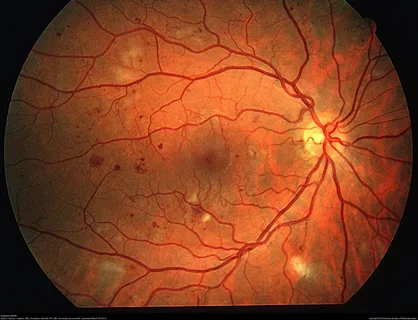
A more serious complication of diabetes affects the back of the eye and can lead to severe visual loss from swelling in the retina. Leakage of fluid or blood from the retinal vessels can progress, leading to hemorrhages and even retinal detachment. What your doctor sees in the retina closely mimics the appearance and function of the vessels in your kidneys and feet.

HYPERTENSION

High blood pressure also causes damage to the retinal vessels. Mild to moderate hypertension is characterized by changes in the vessels that are similar to those seen in early diabetic retinopathy. More acute rises in blood pressure can cause swelling of the optic nerve and a sudden change in vision in one or both eyes.

STROKES

There are several strokes that can directly affect vision. Blood flow through any of the vessels that supply the eye can be disrupted by blood clots, inflammation, or vasospasm. Risk factors for retinal artery or vein occlusions include older age, smoking, high blood pressure, diabetes, obesity, cardiovascular disease and clotting disorders, including malignancy.

Stokes can also affect any of the six muscles that move the eyes up, down, left and right, causing a sudden onset of double vision.
Stokes affecting any part of the visual pathway from the optic nerve to the visual processing centers in the brain can cause a sudden painless loss of vision in one or both eyes. This may affect the entire field of vision or just part of the vision in one or both eyes.

AUTOIMMUNE DISEASES

Many autoimmune diseases like rheumatoid arthritis, lupus, polymyalgia rheumatica and colitis are diagnosed because of ocular manifestations. Iritis, or uveitis, is an inflammatory condition that can affect all layers of the eye. Symptoms may include pain, redness, light sensitivity, decrease in vision and floaters. Some people may be entirely asymptomatic. Treatment includes treating the inflammation and pressure in the eye along with systemic medications prescribed by a primary care doctor or rheumatologist to treat the underlying condition. Complications of these conditions can also lead to glaucoma and cataract formation.

INFECTIONS

Bacterial, viral, fungal and parasitic infections can cause inflammation inside the eye that can mimic auto-immune uveitis. Tuberculosis, Lyme disease, CMV (mono), Herpes Zoster (Shingles) Herpes Simplex, Syphilis, Toxoplasmosis, Histoplasmosis, HIV and several others can cause ocular inflammation with subsequent loss of vision

CANCERS

Several cancers such as breast, lung and prostate cancer tend to metastasize to the eye. Many are detected on routine eye exams before symptoms occur. Primary ocular tumors such as melanoma can be detected by a dilated eye exam. Basal cell, squamous cell and other pre-cancerous lesions have a predilection for the eyelids. Leukemias and lymphomas often present with ocular findings.

MEDICATIONS

Numerous systemic medications cause damage to the eyes and may require baseline screening and regularly scheduled eye exams with ancillary testing to detect signs of toxicity. Plaquenil (hydroxychloroquine) used for rheumatologic diseases, can cause damage to the macula. Yearly dilated exams and visual field testing are needed to screen for irreversible visual loss. Amiodarone (atrial fibrillation) and Ethambutol (TB/MAC) can cause damage to the optic nerve and require regular monitoring by an ophthalmologist. Topamax used for migraines and weight loss, can cause a sudden onset of angle closure glaucoma. Even common medications such as doxycycline, sulfa and hormone replacements can cause a condition that mimics signs of a brain tumor causing headaches, dimming of vision and visual field loss.

Macular Degeneration (AMD) - Amy Cunnigham Eye Consultant

Macular Degeneration (AMD)

WHAT IS MACULAR DEGENERATION?

Age related macular degeneration (AMD) is a deterioration that occurs in the macula, the part of the retina responsible for central vision. AMD is another cause of irreversible vision loss in the aging population. Vision loss typically affects both eyes but at different rates. Because macular degeneration affects central vision, symptoms include difficulty reading, recognizing familiar faces and seeing fine details. There are two forms of AMD: wet and dry. The dry form is more common and visual changes are typically slowly progressive and less severe. The wet form occurs less frequently but with a more sudden onset and more severe visual loss.

WHAT ARE THE RISK FACTORS FOR AMD?

While we don’t know the exact cause of macular degeneration, we can identify several risk factors for developing the disease such as advanced age, smoking, fair pigment (blonde hair/blue eyes) and genetic predisposition. While many of these are not within our control, it has been found that diet and nutrition may play an important role in AMD.

HOW DO I KNOW IF I HAVE MACULAR DEGENERATION?

Some people are diagnosed with AMD after a noticeable decline in vision. Others are diagnosed with early signs of macular degeneration during a routine eye exam. Only a comprehensive dilated eye exam by a medical doctor can detect the early signs of AMD.

CAN VITAMINS HELP PREVENT AMD?

Some patients with AMD may benefit from an over-the-counter formulation that may slow the progression of the disease, but it depends on the type and stage of the disease you have. High levels of specific vitamins and minerals may significantly reduce the progression of AMD and severe vision loss. These supplements may change the way you digest food and may also affect the way your body metabolizes other medications. Therefore, it is always best to talk with your doctor before starting any supplements.

IS THERE A CURE FOR AMD?

While there is no cure for the vision loss associated with AMD there are several new and exciting treatments available to help halt disease progression in selected patients. It is important to see your ophthalmologist to determine if you may be at risk for AMD and to educate yourself on how to detect signs of progression so that treatment can be initiated early on if needed.

WHAT CAN I DO TO STOP VISION LOSS FROM MY AMD?

Early detection of AMD with regular eye exams can help detect any change that may warrant treatment. You can minimize your risk of AMD by not smoking, keeping your blood pressure under control and eating plenty of dark leafy green vegetables and orange fruits and vegetables. Regular home monitoring with an Amsler grid is encouraged.

Download a copy of a real sized Amsler grid from the below link.

http://www.amslergrid.org/

Glaucoma Amy Cunnigham North Shore Eye Consultant

Glaucoma

WHAT IS GLAUCOMA?

Glaucoma is a disease characterized by the buildup of pressure in the eye causing damage to the optic nerve and resultant loss of peripheral vision. The eye is constantly producing and draining fluid internally. Both over production and under filtration of this fluid can cause glaucoma. The optic nerve is made up of about 1 million nerve fibers that carry information from the eye to the brain, allowing us to form images. As these fibers die you develop blind spots in your vision that can easily go undetected for years without formal testing. Glaucoma is one of the leading causes of blindness in people over 60, therefore early detection and treatment are key.

There are several types of glaucoma, the most common of which is chronic open-angle glaucoma. This is characterized by a slowly progressive, painless loss of vision that starts peripherally. It often goes unnoticed by the patient until more advanced disease affects central vision. At this point, it may become dangerous to drive. Although you may see a car directly in front of you, you may have trouble detecting a car in the adjacent lane or a bicyclist or pedestrian crossing the street. The insidious nature of this disease causes irreversible damage if not detected and treated early.

A less common type is acute narrow-angle or angle-closure glaucoma. This is an emergent situation caused by a rapid rise in pressure inside the eye which can cause permanent visual loss within 6 hours. This occurs in predisposed eyes when the angle where the drainage system is located is suddenly compromised, either by cataract progression or the use of numerous common medications. Symptoms of an acute attack of angle-closure glaucoma include severe eye pain or brow ache, redness, blurry vision, halos around lights, nausea and vomiting. This is an emergent situation that requires immediate intervention. Routine eye exams can detect whether you may be at risk for this form of glaucoma. Your doctor may recommend preventative laser treatment or cataract surgery.

WHAT ARE THE RISK FACTORS FOR GLAUCOMA?

While elevated pressure in the eye is one risk factor for glaucoma, there are several others advanced age, family history, race, past ocular trauma, thin corneas and near-sightedness. Some people develop glaucoma with “normal eye pressure”. Several systemic diseases including diabetes, hypertension, migraines, sleep apnea and anemia are predisposing factors.

HOW DO I KNOW IF I HAVE GLAUCOMA?

Routine screening eye exams are the best way to detect glaucoma. It is important to understand that the most common type of glaucoma has no symptoms until the disease is quite advanced. Because over half of the nerve fibers can die before there is a detectable change in vision, we use highly magnified lenses and three-dimensional imaging techniques to examine your optic nerve along with formal visual field testing to detect glaucoma in the earliest stages and monitor disease progression. Our office is equipped with the latest technology to assess, treat and monitor glaucoma.

WHAT IS THE TREATMENT FOR GLAUCOMA?

Treatment for glaucoma is aimed at lowering the pressure inside the eye to prevent further visual decline. There is no cure for glaucoma and no treatment that will restore vision once it is lost, therefore early detection and treatment are important.

In open-angle glaucoma, pressure can be reduced by various methods including eye drops, laser procedures and surgery in the operating room. Some patients achieve adequate pressure reduction with eye drops alone. Some patients are better candidates for laser treatment. SLT stands for selective laser trabeculoplasty. It is a painless procedure that is done in the office to help reduce pressure in the eye. Many patients require both topical and laser treatment. Few patients undergo surgery.

In narrow-angle glaucoma, treatment may initially be eye drops, followed by a laser procedure called a peripheral iridotomy. The YAG laser makes a small hole in the iris, the colored part of the eye so that pressure will not build up inside the eye. This hole is NOT a connection to the outside of the eye and is not visible to the naked eye. Our office has the latest state-of-the-art equipment to both diagnose and treat both forms of glaucoma.

CAN MY GLAUCOMA BE CURED?

There is no cure for glaucoma and neither medicine nor surgery will restore vision once it is lost. Only a comprehensive eye exam and evaluation of your individual risk factors can detect glaucoma early. You and your doctor will determine when treatment should be initiated and which treatment option is right for you.

Cataracts Amy Cunnigham North Shore Eye Consultants eye Services

Cataracts

WHAT IS A CATARACT?

The eye has a lens, like a camera, that focuses rays of light onto the retina. A cataract is a hardening or clouding of the eye’s natural crystalline lens. Cataracts develop as part of the normal aging process, causing progressive blurry central vision, glare and halos when driving at night. They can also be caused by certain disease processes or by the medications used to treat those conditions. Because cataracts change the way that light is bent through the lens and focused on the retina, early cataracts can often be addressed simply by changing your glasses prescription. Once vision is compromised and you are no longer able to do the things you enjoy, cataract surgery should be considered.

HOW IS CATARACT SURGERY DONE?

Dr. Cunningham performs modern day small incision cataract surgery at Glenbrook Hospital. The surgery is done under a microscope with topical anesthesia and IV sedation. A small incision is made at the edge of the cornea. It is a common misconception that a laser is used to remove a cataract. Instead, ultrasound is used to break up the cloudy lens into small pieces that are then aspirated from the eye. A permanent intraocular lens of the appropriate pre-determined power is then implanted to replace the natural lens. In most cases, the original incision is small enough that it self-seals and requires no sutures. The procedure typically takes about 15 minutes and is done on an outpatient basis, so you go home the same day with minimal restrictions. Most patients report no discomfort during cataract surgery, just an interesting “light show.” After surgery, a few patients feel a mild scratchy sensation which typically resolves on its own.

CAN CATARACTS RECUR AFTER SURGERY?

Cataracts cannot recur after surgery because the natural lens has been removed. In about 30% of patients, the capsule that holds the implant in place can become cloudy over time. This is called a “secondary cataract”. If vision is compromised by this scar tissue, a YAG laser is used to make an opening in the capsule. It is a painless in office procedure that takes less than 5 minutes with an immediate improvement in vision.

WHEN IS IT TIME TO REMOVE MY CATARACTS?

In years past, we waited until your cataracts were “ripe.” before removing them. This is no longer considered the standard of care. Advances in surgical techniques and the risk of complications that come with removing dense cataracts have made it possible to remove them earlier, without a major disruption to your lifestyle. You, as the patient, will be very involved in making that decision. If your vision is interfering with your activities of daily living and cannot be improved to your satisfaction with glasses, then it is time to consider surgery. Only a comprehensive exam and detailed discussion with your doctor can guide you.

PREMIUM INTRAOCULAR LENSES

Selecting the right implant can be an important decision for many people undergoing cataract surgery. Measurements are taken before surgery to determine what power lens implant you will receive. In the past, most patients required glasses or bifocals after surgery in order to see to their satisfaction. This is no longer the case with modern day premium intraocular lens implants.

Imagine the ability to drive, cook and read without glasses! Dr. Cunningham offers a variety of modern intraocular lens implants to reduce your dependence on glasses altogether. Many lens implants are designed to allow you to have good distance, intermediate and near vision without the strong dependence on glasses associated with traditional lens implants. Advances in technology also allow for the correction of long-standing astigmatism caused by an abnormal curvature of the cornea. Many people enjoy the vision they experienced in their 30’s without glasses! Talk with Dr. Cunningham to determine if you are a candidate for one of these innovative implants.